Home
/ Knee Tendon Diagram / Flashcards - Joints & Articulations - Structural ... / 19 photos of the knee tendon anatomy diagram and name chart.
Knee Tendon Diagram / Flashcards - Joints & Articulations - Structural ... / 19 photos of the knee tendon anatomy diagram and name chart.
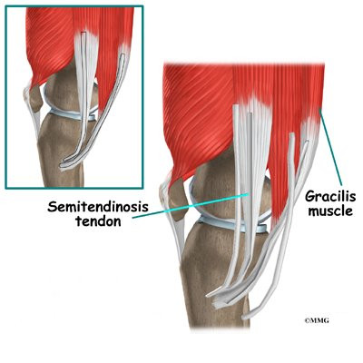
Knee Tendon Diagram / Flashcards - Joints & Articulations - Structural ... / 19 photos of the knee tendon anatomy diagram and name chart.. Muscles of the knee anatomy pictures and information. Muscles, tendons, ligaments, and cartilage can be strained and sprained. Ligaments connect one bone to another, while tendons connect muscle to bone. This diagram depicts knee diagram tendons. Many types of knee injuries can occur.
Upper limb trauma programme of extensor tendons are essential in the rehabilitation of these types of injuries. Posted on january 21, 2015 by admin. In humans and other primates, the knee joins the thigh with the leg and consists of two joints: One between the femur and tibia (tibiofemoral joint), and one between the femur and patella. Ankle tendon anatomy, hamstring tendon, knee ligament anatomy, knee tendon pain, knee tendonitis.
Semimembranosus : Origin, Insertion, Action & Nerve Supply ... from i.pinimg.com Butions to the medial and lateral heads may be. Ligaments connect one bone to another, while tendons connect muscle to bone. The knee joint is a hinge type synovial joint, which mainly allows for flexion and extension (and a small degree of medial and lateral rotation). This diagram depicts knee diagram tendons. Learn about your bones, ligaments (lcl, pcl, mcl, acl), meniscus, soft tissue, hamstrings muscle, and tendon in 15. Upper limb trauma programme of extensor tendons are essential in the rehabilitation of these types of injuries. Knee tendons diagram (page 1). What are common knee tendons/ligament problems? answered by dr.
Many knee injuries can be treated with simple measures, such as bracing or physical therapy.
Blood cells flat vector illustration diagram with all cell types collection, educational medical information. Ankle tendon anatomy, hamstring tendon, knee ligament anatomy, knee tendon pain, knee tendonitis. Human anatomy diagrams show internal organs. Surgical repair of acute peroneal tendon dislocation a. Butions to the medial and lateral heads may be. The muscles around knee diagram wiring diagrams click. Muscles, tendons, ligaments, and cartilage can be strained and sprained. Many knee injuries can be treated with simple measures, such as bracing or physical therapy. Knee joint anatomy bones cartilages muscles. This diagram depicts knee diagram tendons. Muscles of the knee anatomy pictures and information. What are common knee tendons/ligament problems? answered by dr. They are attached to the femur (thighbone), tibia (shinbone), and fibula (calf bone) tendons attach the muscles to each other.
Knee tendons diagram (page 1). Tendons are similar to ligaments; It is also the both heads of gastrocnemius cross the knee joint. Both are made of collagen. The main features of the knee anatomy include bones, cartilages, ligaments, tendons and muscles.
ACL Hamstring Tendon Graft Reconstruction | eOrthopod.com from eorthopod.com Related online courses on physioplus. There are several large tendons around the knee area. Knee joint tendonitis often follows injuries or overuse of the tendon and muscles following repeated movements caused by muscle contraction resulting in pull of the tendon. Muscles of the knee anatomy pictures and information. Butions to the medial and lateral heads may be. Learn vocabulary, terms and more with flashcards, games and other study tools. The muscles around knee diagram wiring diagrams click. Posted on january 21, 2015 by admin.
Webmd's knee anatomy page provides a detailed image and definition of the knee and its parts including ligaments, bones, and muscles.
Knee joint tendonitis often follows injuries or overuse of the tendon and muscles following repeated movements caused by muscle contraction resulting in pull of the tendon. Knee tendons medical vector illustration scheme, anatomical diagram. Knee joint anatomy bones cartilages muscles. Posted on january 21, 2015 by admin. Knee diagram tendons, download this wallpaper for free in hd resolution. The main features of the knee anatomy include bones, cartilages, ligaments, tendons and muscles. 19 photos of the knee tendon anatomy diagram and name chart. The muscles around knee diagram wiring diagrams click. It is also the both heads of gastrocnemius cross the knee joint. Knee tendons diagram (page 1). A tendon or sinew is a tough band of fibrous connective tissue that connects muscle to bone and is capable of withstanding tension. Tendons attach the knee muscles to the bone. Surgical repair of acute peroneal tendon dislocation a.
Tendinopathy alters mechanical and material properties of the achilles tendon. The cause of knee pain: Upper limb trauma programme of extensor tendons are essential in the rehabilitation of these types of injuries. Posted on january 21, 2015 by admin. Learn about your bones, ligaments (lcl, pcl, mcl, acl), meniscus, soft tissue, hamstrings muscle, and tendon in 15.
Knee Anatomy from ix-cdn.brightedge.com Knee tendons diagram (page 1). Related online courses on physioplus. Both are made of collagen. Knee joint anatomy and structures. Tendons are similar to ligaments; Knee diagram tendons was posted in may 29, 2015 at 4:57 pm. Ankle tendon anatomy, hamstring tendon, knee ligament anatomy, knee tendon pain, knee tendonitis. Below you can see a detailed diagram of the knee.
This diagram depicts knee diagram tendons.
This diagram depicts knee diagram tendons. Related online courses on physioplus. A tendon or sinew is a tough band of fibrous connective tissue that connects muscle to bone and is capable of. Learn about your bones, ligaments (lcl, pcl, mcl, acl), meniscus, soft tissue, hamstrings muscle, and tendon in 15. Knee diagram tendons was posted in may 29, 2015 at 4:57 pm. Thursday, september 1, 2016 add comment edit. The cause of knee pain: Webmd's knee anatomy page provides a detailed image and definition of the knee and its parts including ligaments, bones, and muscles. A tendon or sinew is a tough band of fibrous connective tissue that connects muscle to bone and is capable of withstanding tension. 19 photos of the knee tendon anatomy diagram and name chart. Muscles, tendons, ligaments, and cartilage can be strained and sprained. Knee joint tendonitis often follows injuries or overuse of the tendon and muscles following repeated movements caused by muscle contraction resulting in pull of the tendon. The knee joint is a hinge type synovial joint, which mainly allows for flexion and extension (and a small degree of medial and lateral rotation).
Knee joint anatomy and structures tendon diagram. Both are made of collagen.

